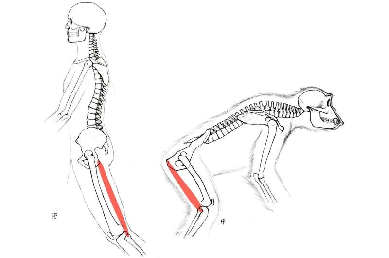
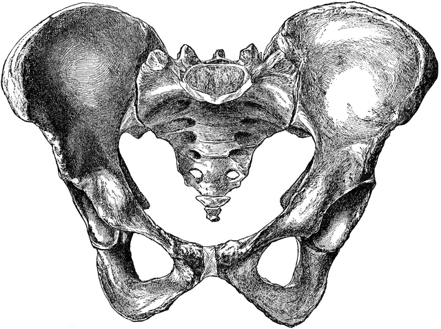
One distinguishing physical characteristic of our species is the broad, basin-shaped human pelvis. Without it, we wouldn’t be able to stand up straight or have children with large brains. New research on human embryos has now pinpointed the developmental stage at which the pelvis starts to resemble a human and named hundreds of genes and regulatory RNA regions responsible for this change. The authors draw the conclusion that many exhibit signs of strong natural selection for bipedalism.
According to Marcia Ponce de León, an anthropologist at the University of Zürich “It is a really impressive study, especially the genomic part, which uses all the bells and whistles of state-of-the-art-analysis,” The findings, she continues, are consistent with the notion that evolution frequently generates novel physical traits by influencing genetic switches that have an impact on early embryonic development. It is “easy to state but very difficult to actually demonstrate, and this is what the authors did,” that such predictions are.
According to the article published in Science by Michael Price, in primates, the pelvic girdle is made up of three main components: the pubis and ischium, which form the shape of the birth canal; the ilia, which have a blade-like shape and fan out to form the hips. Great apes have birth canals that are relatively narrow and ilia that are relatively elongated and lie flat against the animals’ backs. The ilia of humans are rounded, shorter, and they flare out and curve around. Our large-headed newborns can fit through a wider birth canal thanks to the reshaped ilia, which also serve as attachment points for the muscles that stabilize upright walking. According to Terence Capellini, an evolutionary biologist at Harvard University, those pelvic patterns were already developing in 4.4 million years old Ardipithecus ramidus, a hominin with slightly turned-out ilia and suspected occasional two-footed activity.
However, it had been unclear as to when and how those characteristics developed during human gestation. By week 29, many of the crucial aspects of the human pelvis, such as its curved, basin-like shape, are already formed. However, Capellini questioned whether they might appear earlier, when the cartilage scaffolding of the pelvis still exists and the pelvis has not yet turned to bone.
The researchers examined 4- to 12-week-old embryos under a microscope with the consent of women who had legally terminated their pregnancies. They discovered that the ilium starts to form and rotate into its distinctive basin-like shape around the 6- to 8-week mark. Capellini’s team discovered that this cartilage stage in the pelvis appears to persist for a number of additional weeks, giving the developing structure more time to curve and rotate, even as other cartilage within the embryo starts to ossify into bone. “These aren’t bones, this is cartilage that is growing and expanding and taking that shape,” Capellini says.


The next step was to extract RNA from various pelvic regions of the embryos to determine which genes were active at various developmental stages. After that, they discovered a large number of human genes within particular pelvic sections whose activity appeared to change during the first trimester. 261 of these genes were located in the ilium. According to Capellini, many of the upregulated genes maintain cartilage while many of the downregulated genes are involved in converting cartilage to bone. This may help keep the ilium in a cartilaginous stage for longer.
The researchers also discovered thousands of genetic on/off switches that appeared to be involved in forming the human pelvis by contrasting the genetic activity of the developing pelvis with that of a mouse model. Since our species diverged from the chimpanzees that we share a common ancestor with, DNA stretches within those switches seem to have undergone a rapid evolution. However, those regulatory regions of the ilium exhibit remarkably little variation in modern humans. According to the researchers, this uniformity is evidence that the ilium has been under intense pressure from natural selection to develop in a very specific manner.
Capellini “We think this is really pointing to the origins of bipedalism in our genome,” said.
The evidence for strong selective pressure on the pelvis is not surprising to UZH anthropologist Martin Häusler, but he adds that the results provide an impressive look into some of the pelvic changes “at the very origin of what makes us human.” He saying that rather than using mouse models, future research comparing human embryos to those of other primates would provide a better understanding of how natural selection reshaped the human pelvis.
According to Nicole Webb, a paleoanthropologist at the University of Tübingen who studies pelvic anatomy, the growing understanding could also aid researchers in developing treatments for hip joint conditions or forecasting complications during childbirth. Deviations from the genetic program that Capellini’s team identified can result in disorders such as hip dysplasia and hip osteoarthritis, she says. “I hope that it has major implications for making people’s lives better. That would be huge, to connect paleoanthropology with real life.”
